
Background
Women are two to ten times more likely to develop a fatigue-induced fracture whether from osteoporosis, military combat training, or sports. This increased risk has long been thought to be a consequence of intrinsic factors such as smaller bones, lower bone mineral density (BMD), lower cortical thickness, hormonal imbalance, etc. However, a robust fracture-risk assessment has yet to be developed using these predictors, and stress fractures remain a tremendous economic burden and cause of morbidity. Since stress fractures most likely occur due to over-accumulation of unrepaired bone microdamage, men and women’s bones may develop and/or respond differently to microdamage. If so, a sex-specific understanding of 1) bone quality, 2) microdamage development with fatigue loading, and 3) the correlation between the amount of microdamage and bone quality degradation would significantly advance our understanding of stress fractures.
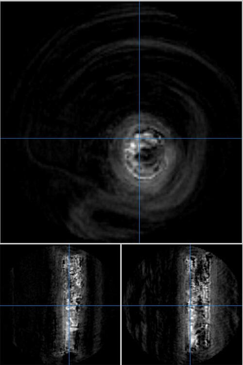
Figure 1: Axial (top) and sagittal (bottom) images of water content in a sample of cortical bone. Brighter areas are saturated with more water compared to darker areas of the image.
The most clinically applicable advancement in this space would entail the assessment of microdamage in vivo. As such, this study proposed to achieve a key advancement of in vivo microdamage assessment using magnetic resonance (MR) imaging. Microcracks are essentially “pores” in which water can pool as unbound water. Therefore, we hypothesized that the amount of microdamage that occurs due to fatigue loading could be measured using 3D ultrashort echo time cones MR imaging and correlated to standard microdamage quantification techniques. This capability would provide a means to assess microdamage in vivo and enable new fracture risk prediction methodologies. To this end, the goal of this project was to develop an MRI technique that is capable of assessing water content at the microscale in bone.
Approach
Two frozen baboon femurs were cut to isolate each diaphysis. The bones were loaded in 3-point bending. An imaging protocol was developed to identify unbound water in bone with high resolution and medical physicists from UT Health implemented the protocol and imaged the loaded bones.
Accomplishments
The conclusion of this program resulted in the ability to image water in bone (Figure 1) where bright areas of the image data indicate the presence of unbound water. With this newly developed ability to image water in bones at a high resolution, future work and external proposals will leverage this capability and perform larger-scale studies to assess water changes before and after loading as a means to quantify microdamage in bone.