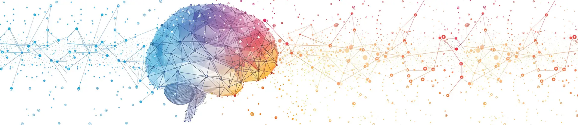
The brain is the most critical and complex organ in the human body. Three pounds of gray matter nestled in the human skull are the source of our intelligence, senses, movement and behavior — the qualities that define our humanity.
In 2024, Southwest Research Institute launched a brain health working group to bring together multidisciplinary specialists studying everything from how neurons grow and connect, to screening and preventing brain injuries and assessing decline.
The group’s goal is to create multifunctional and multidisciplinary tools and resources at SwRI to support brain health research and development. The team wants to build platform-based technology that uses machine learning to create tools using data from multiple research initiatives and sources. Examples include traumatic brain injury (TBI) screening tools and techniques, sensor systems and various measurement and diagnostic tools. The goal is to combine objective data to find patterns across tools, integrating with electronic charting and patient tracking systems to provide a continuity of care while continuing to evolve and improve through expanded data learning algorithms.
The team aims to provide an adaptable platform that provides accessible, relevant and understandable information quickly and reliably through all phases of care.
This listicle outlines just a few projects underway across SwRI. For instance, machine learning specialists are looking at how neuronal networks form in the brain and how that could affect brain health as well as understanding how cognitive load, the amount of mental effort needed to perform a task, affects function. Chemists are looking at new and creative ways to screen for TBIs that are less expensive and invasive, while mechanical engineers are looking at high-tech helmets designed to prevent injury. And human performance engineers are looking at how to detect cognitive decline using the kinematics of gait.
Research into TBIs is a particular focus, in part because the effects vary so widely, from minor to lifechanging and everything in between. And TBIs — caused by a forceful bump, blow or jolt to the head or body or from an object entering the brain — are common. In the U.S., TBIs result in 200,000 hospitalizations and 70,000 deaths a year. Some TBIs cause temporary or short-term problems with brain function, including problems with cognition, comprehension, movement, communication and actions. More serious TBIs can lead to severe or permanent disability and even death. Causes include falls, motor vehicle crashes, sports injuries, blasts, battlefield trauma or blows by an object.
Explore the following six targeted research programs making waves in brain research.
1. NEURAL CONNECTIONS
Understanding how neurons evolve and form networks in the brain could lead to new drug treatments for people suffering from neurological disorders. A team led by SwRI developed a computational method to improve techniques to track brain cell development over time.
![]()
Dr. Courtney Rouse, a senior research engineer in SwRI’s Intelligent Systems Division, led research to develop a computational method to improve techniques to track brain cell development over time.
SwRI collaborated with The University of Texas at San Antonio (UTSA) to track individual brain cells undergoing neurogenesis, the process where new neurons grow and connect to neuronal networks. UTSA researchers developed novel methods to grow human stem cells that form networks, such as those that regulate sleep, temperature and mood. As the neuronal networks developed, their activity was captured by confocal microscopy. SwRI then used the visual data to train algorithms to track the neurons. In the future, these algorithms could automatically classify the health of growing neuronal networks to study various neurological diseases and help develop and test associated therapies.
Conventional neuron-tracking methods frequently rely on images of labeled cells in fixed tissue, a process that can obscure cellular dynamics. The SwRI-led analysis overcomes technological gaps in prior computational methods by capturing unlabeled cells and fine structures in dense, live cultures. This allows for complex analysis over longer time spans. Each video of the neuronal network cultures consists of timestamped images with hundreds to thousands of neurons per image. SwRI trained a U-Net machine learning algorithm to detect the shape and location of individual neurons in the images.
SwRI trained the algorithm to recognize two of the main parts of a neuron — the soma, which contains the nucleus, and dendrites, root-like structures connecting with other neurons, forming neuronal networks that send and receive signals.
The team focused on tracking the somas, because each neuron has one soma, while dendrite numbers vary. The algorithm applied a tracking number to a key point on each soma to match neurons in consecutive images based on proximity within a pixel. When detecting somas, the algorithm achieved a 93.8% precision rate and a 99.1% recall rate.
When detecting dendrites, the smaller branch-like structures, the algorithm achieved an 88.3% precision rate and an 80.9% recall rate. Overall, the algorithm had an 85.7% probability of correctly tracking a single neuron in consecutive images.
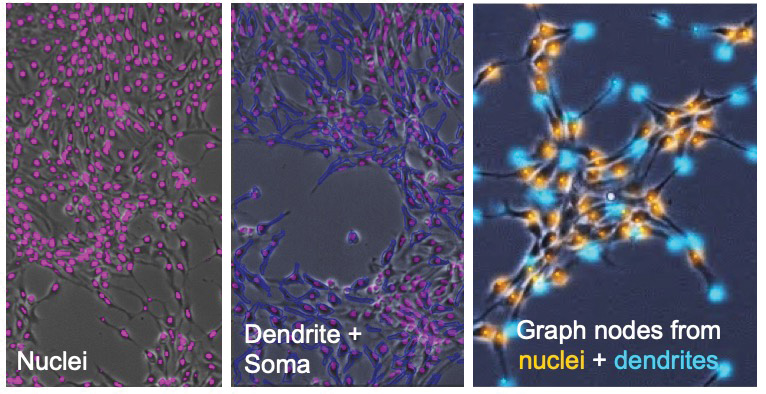
Using UTSA’s video data of developing neurons, SwRI trained various algorithms to detect neurons and their components. From left, the team trained a U-Net algorithm to detect somas (pink) and an instance-aware semantic segmentation algorithm to detect whole neuron contours (blue) overlaid on the somas (pink). They used a convolutional neural network based on Inception V3 to detect somas and dendrites.
The team plans future research to identify connections between soma and dendrites, test neurons exposed to various environmental stresses — such as low oxygen or circadian disruption — and correlate neuron electrical activity to tissue health.
The research helped bridge technological gaps in computational neural research. The algorithm accurately tracked individual soma across timeframes, a fundamental step toward classifying the health of a developing neuronal network. The project was funded by a \$200,000 award from the San Antonio Medical Foundation.
Questions about this story and Bioinformatics Data Analysis Services? Contact Dr. Courtney Rouse at + 1 210 522 6873.
2. COGNITIVE LOAD
Cognitive load refers to the mental effort needed to process information, and consequently, excessive loads can hinder learning and performance. For example, a student struggling to solve a complex math problem while simultaneously learning the underlying concepts for the first time illustrates high cognitive load.
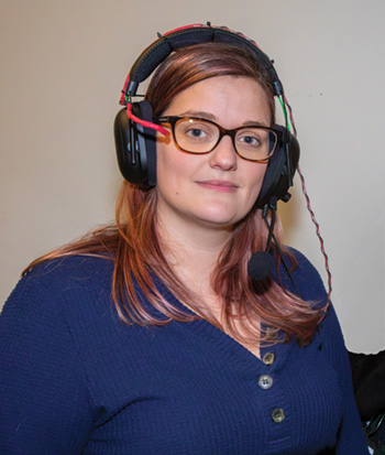
Dr. Katelyn Fry-Hilderbrand, a research engineer in SwRI’s Intelligent Systems Division, developed a fieldable system to assess cognitive performance in diverse settings.
The concept of cognitive load is central to understanding how the brain allocates its working memory resources, vital for generating thoughts and solving problems. When overwhelmed, cognitive overload hampers the ability to think, learn, remember and solve problems. The ability to assess cognitive load is essential to some professions, particularly for the military, surgeons, long-distance drivers and welding, to name a few.
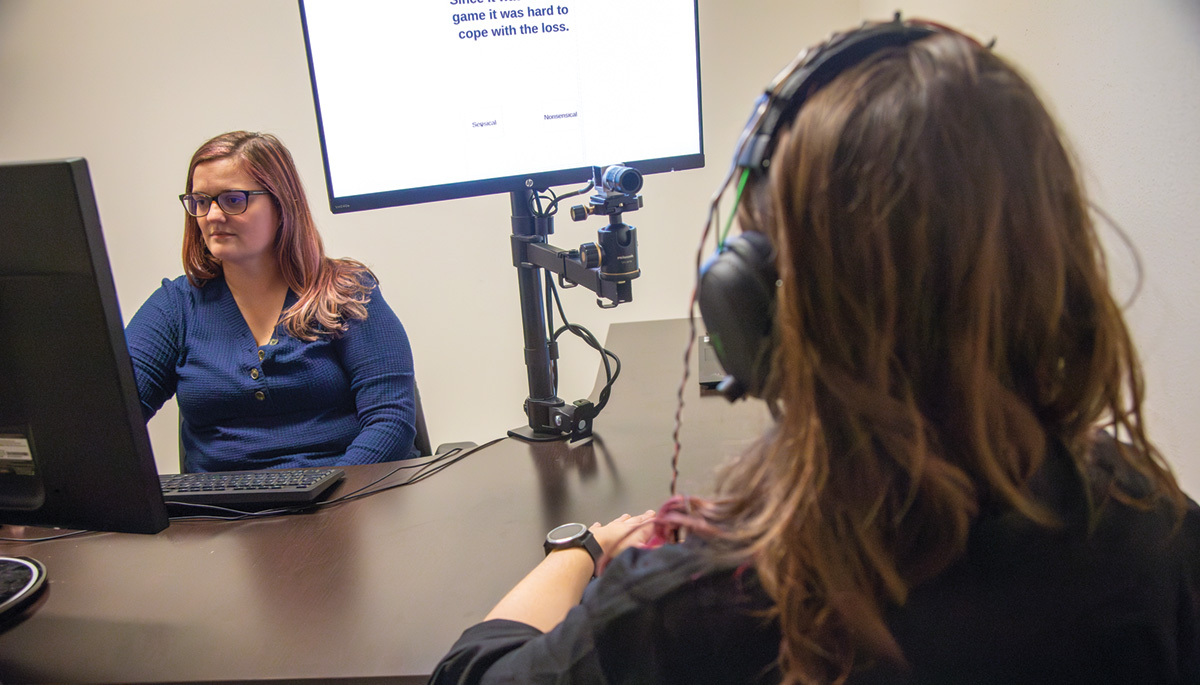
As part of an internal research program, Southwest Research Institute explored psychophysiological measurements to assess cognitive load, aiming to create a field-ready cognitive load assessment tool. Traditionally, getting these measurements required controlled environments, limiting their broader applicability. SwRI created a data collection system using off-the-shelf components to assemble data similar to those gathered by traditional lab-based systems, including electrocardiography sensors, voice recorders and eye-tracking cameras.
Using this system, subjects underwent tasks designed to increasingly challenge cognition until they reached an overloaded state. The team then trained predictive models using data from both setups to predict cognitive performance and compared the accuracy of these models to validate the fieldable system. Models trained using the traditionally gathered dataset and the fieldable dataset had similar predictive powers, validating the use of the fieldable system.
Fry-Hilderbrand explored psychophysiological measurements to assess cognitive load, which is central to understanding how the brain allocates its working memory resources and vital for generating thoughts and solving problems.
Additionally, the team found that individualized models improved accuracy significantly compared to generalized models. Larger training datasets could enhance the accuracy of general models, while individualized models promise better predictions with fewer samples. The successful implementation of the fieldable system enables SwRI to assess cognitive performance in diverse settings.
As part of a continuing research effort, the team used the fieldable system to analyze the impact of various stressors on cognitive performance, including how cognitive performance changed with the introduction of physical stress. The team evaluated cognitive performance before and after physical loading tasks to understand the impact of physical exertion. Analysis revealed a general decline in cognitive performance after physical tasks, though not uniformly, suggesting a nuanced relationship between physical load and cognitive capability, where moderate physical exertion might benefit cognitive performance. Upcoming research will examine the relationship between cognitive performance and other stressors such as fatigue, emotional stress, etc.
Questions about this story and Bioinformatics Data Analysis Services? Contact Hakima Ibaroudene at + 1 210 522 3963.
3. TBI SCREENING KIT
Traumatic brain injuries (TBIs) can have profound and sometimes long-term effects that can dramatically change the course of lives. They are also notoriously difficult to diagnose, and multiple TBIs can be catastrophic.
Kreg Zimmern, a senior research engineer in the Chemistry and Chemical Engineering Division, championed the AMMO field-ready TBI screening kit, with potential uses from the battlefield to the football field. The test identifies the inability to smell, which correlates highly with a TBI.
To address this, SwRI developed a field-ready screening tool for TBIs. The Advanced Military Measure of Olfaction (AMMO) kit includes an array of scents, deployable anywhere from the battlefield to the football field, to help screen for TBIs in minutes.
The AMMO test kit is not intended as a diagnostic test but as a screening tool. The kit includes six sealed vials that release a range of odors, such as fruity and spicy aromas. When squeezed, a test vial turns blue to indicate a smell has been released and is ready for use. Patients are asked to identify the odor from four possible choices on an attached card. Answers are documented onto a separate answer card with the correct answers hidden behind a sticker. Research indicates that failing to identify the scents correctly correlates highly with positive results for TBI on an MRI exam.
Someone exposed to a blast on the battlefield could be screened immediately with AMMO instead of waiting for the onset of signs or symptoms of TBI. The inability to identify the scents could be used as rationale to justify an MRI. Similarly, someone in a rural area or at a sports tournament could be AMMO tested to determine if a trip to an emergency room is warranted.
AMMO is currently undergoing stability studies to determine how long the kit can be stored and remain effective. SwRI is developing AMMO in compliance with relevant Food and Drug Administration and ISO guidelines. It’s the only olfactory test kit to undergo such rigorous controls.
The kit is inexpensive, compact, has no special storage conditions and doesn’t need electricity. This makes it a potential screening tool in emergency rooms as well as at workplaces, nursing homes and youth, collegiate and professional sports games. While AMMO doesn’t require any specialized training to administer, the results can inform the decision-making process for first responders and doctors.

The AMMO kit includes six sealed vials that release a range of odors, such as fruity and spicy aromas. Research indicates that failing to identify the scents correctly correlates highly with positive results for TBI on an MRI exam. When squeezed, a test vial turns blue to indicate a smell has been released and is ready for use. Patients are asked to identify the odor from four possible choices on an attached card.
SwRI collaborated with the Henry M. Jackson Foundation for the Advancement of Military Medicine and the Air Force Research Laboratory to develop AMMO.
Questions about this story or Biochemistry & Bioengineering? Contact Kreg Zimmern at +1 210 522 2493.
The views expressed are those of the authors and do not reflect the official guidance or position of the United States Government, the Department of Defense or the United States Air Force. The contents of this publication are the sole responsibility of the author(s) and do not necessarily reflect the views, opinions or policies of The Henry M. Jackson Foundation for the Advancement of Military Medicine, Inc.
4. BREATH-BASED BIOMARKERS
Globally, traumatic brain injuries (TBIs) affect approximately 50 million people and are a leading cause of cognitive and behavioral decline. To address this pressing issue, researchers are striving to develop diagnostic tools that are rapid, sensitive and accurate. SwRI is exploring the use of metabolic biomarkers in breath to create a noninvasive diagnostic solution for TBIs as part of its Brain Health Working Group.
Dr. Mark Libardoni, a staff scientist in the Space Science Division, has been working on breath-based biomarkers as part of SwRI’s Brain Health Working Group. Libardoni has developed novel methodology that allows organic compounds in breath to be analyzed by gas chromatography and mass spectrometry.
Human breath is abundant with organic compounds that form an individual’s unique “breathprint.” This breathprint reflects the levels of metabolites — small molecules like amino acids and lipids — produced through the body’s chemical processes. Breath analysis, a practice rooted in ancient Chinese medicine, has more recently been employed to diagnose and manage diseases such as lung cancer, prostate cancer, breast cancer and even Parkinson’s and age-related dementia such as Alzheimer’s. Over the past five decades, advances in analytical instruments and computing have enabled scientists to decode the composition of breath and link it to specific health conditions.
In medical applications, breath analysis can differentiate between healthy and diseased states and monitor health changes over time. It offers remarkable sensitivity and detectability, complementing traditional blood and urine tests while enhancing research capabilities as a non-invasive screening tool.
SwRI collaborated with the University of Texas at San Antonio (UTSA) to identify breath biomarkers linked to mild TBIs, including concussions and sub-concussions. The research involved equipping participants with accelerometer-embedded mouthguards to measure impact forces as they repeatedly headed a soccer ball. Researchers collected breath and blood samples at multiple intervals — before the activity, immediately after and over the following seven days. While UTSA analyzed blood samples for known brain inflammation markers, SwRI examined breath samples for various organic compounds. The data were then integrated and processed using machine learning to pinpoint biomarkers associated with brain inflammation.
SwRI’s expertise in breath analysis began with developing technology to monitor air quality and astronaut breath biomarkers on the International Space Station. For the TBI research, scientists captured organic compounds from breath samples in sorbent tubes, which were thermally desorbed and analyzed in the lab. Gas chromatography- mass spectrometry (GCMS) separates and identifies organic compounds in the sample. Concurrently, materials scientists and engineers worked on miniaturized sensors tailored to detect TBI specific biomarkers in breath.
Breath contains a wealth of information, with upwards of 1,500 detectable compounds. Using GCMS data analysis tools, researchers isolated key compounds based on factors such as concentration and chemical structure. These data were then shared with biostatisticians and modelers to identify the most relevant TBI biomarkers, both visually and numerically.
The ultimate goal is to create portable, handheld diagnostic devices capable of detecting these biomarkers. Such devices would allow users to simply exhale into them, enabling rapid, on-site and highly accurate TBI diagnoses.
Questions about this story or Multi-Dimensional Gas Chromatography? Contact Dr. Mark Libardoni at +1 210 522 2205.
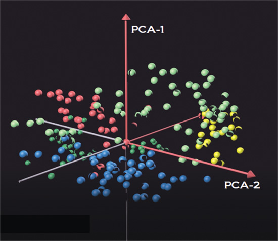
After collection and processing, biostatisticians use mathematical tools to determine the statistical relevance of identified compounds. With hydrocarbons in blue, ketones in yellow, aldehydes in green and fatty acids in red, the biomarkers are represented as a function of their chemical family and amount of variance between healthy and injured states. From here, scientists can determine the compounds specific to TBI.
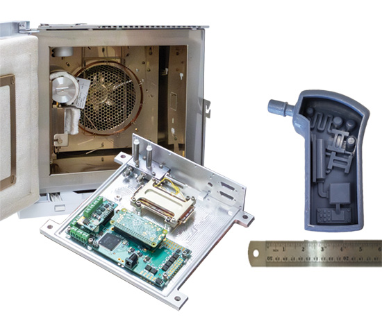
SwRI developed small, rugged technology (right) to perform GCMS (conventional system shown in background left) analyses to monitor the air quality and astronaut breath biomarkers on the International Space Station. The team is now miniaturizing that technology further to create a handheld diagnostic device (right) capable of detecting TBI biomarkers in the field.
5. TBI PREVENTION
Incidents of TBIs are prevalent in military personnel as a result of blast exposures, blunt trauma and ballistic impacts encountered in combat and training scenarios. These profound injuries affect not only the physical health of the service members but also their mental health, cognitive functioning and quality of life.
Dr. Daniel Portillo, a research engineer in SwRI’s Mechanical Engineering Division, is studying how new helmet designs and high-tech pad materials can provide the best protection against battlefield injuries.
Helmets, the primary form of protection against these threats, have undergone significant improvements over the last 70 years. Emerging technologies and materials provide new options for improving a helmet’s ability to reduce the risk of TBI among service members. SwRI is currently focused on the padding within military helmets, where efforts aim to reduce injury by developing pads that adapt to forces applied. For instance, protecting against blunt impact forces needs a softer pad, something like foam, while ballistic impact forces are best mitigated by stiffer pads, like rubber. The goal is to engineer a material that changes its behavior based on the type of impact. The outer shape of the pad and helmet reduces the force of blast impact.
Computational modeling allows engineers to explore novel helmet padding materials and geometries to be optimized for specific cases like blunt, ballistic and blast impacts. SwRI has a history of conducting high-fidelity and experimentally validated simulations for a range of dynamic events. The team uses a variety of software tools to model these events, including the Elastic Plastic Impact Computation, or EPIC, dynamic finite element code. EPIC is managed and distributed by SwRI to model wave propagation, elastic-plastic flow, material failure, fragmentation and complex interfaces.
Over the past 20 years, SwRI has also developed high-fidelity computational models of the human body to investigate numerous aspects of human biomechanics and performance, injury assessment and mitigation, and musculoskeletal disease and joint replacement. Currently, engineers are incorporating a high-fidelity brain model into the existing full-body finite element model. Combining both types of simulations will predict how the helmet pad performs and what level of injury a user would sustain during an actual event.
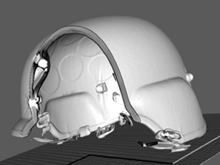
To develop better military helmets, SwRI uses CT imaging to visualize and characterize how composite materials halt ballistic projectiles.
While models are extremely useful in understanding the exact mechanics and interaction between an impact event and the human body, they are often validated and fine-tuned using experimental data. SwRI has decades of experience in the ballistic and explosive testing required to validate the computational models of the helmet pads.
Ballistics tests are often conducted using powder guns or compressed gas guns, which can launch a wide variety of projectiles at a wide range of speeds. Explosive tests are often conducted in blast chambers, at various open-air test sites or replicated using shock wave-generating devices. SwRI uses both types of tests to study how various materials, armors and protection systems respond to impacts. SwRI is combining its experience in cutting-edge materials, computer simulations and experimental validations to rapidly create new helmet pads and other technologies to better protect those who protect us.
Questions about this story or Armor Mechanics? Contact Dr. Daniel Portillo at +1 210 522 4688.

To improve helmet padding materials, Portillo (left) collaborated with the University of Texas at San Antonio’s Morteza Seidi, evaluating military helmet protection against small arms fire.
6. DETECTING DECLINE
Gait analysis, the way a body moves through space, can be used to diagnose neurodegeneration. Subtle shifts in the way we walk can serve as an early indication of cognitive decline. Traditional clinical assessments have relied on basic spatiotemporal measures such as gait speed and stride length gathered from instrumented mats. However, these methods often fall short of capturing the intricate details of motor impairments. The focus on the effects of altered motor control potentially overlooks subtle gait anomalies. Plus, clinical settings can affect natural behavior.
Dr. Dan Nicolella, an Institute Engineer in SwRI’s Mechanical Engineering Division, co-leads the team that developed SwRI’s ENABLE® markerless motion capture technology. Engineers use ENABLE to assess the full range of human performance, including early signs of cognitive decline using biomechanical assessments of gait..
SwRI is applying its ENABLE® markerless biomechanics system to accurately and automatically quantify a person’s detailed 3D movement from video alone, providing a comprehensive full-body gait analysis. It captures kinematic variables in the hips, knees, ankles, shoulders, elbows, etc. The system also captures joint angles, body sway and trunk rotation, offering a far more sensitive assessment of neuromotor dysfunction.
The ENABLE system produces full-body biomechanical analyses from standard video equivalent to laboratory-grade marker-based motion analysis systems. ENABLE offers the same high-fidelity data without the cumbersome markers and specialized, expensive cameras. By measuring full-body biomechanics simultaneously with traditional spatiotemporal data from an instrumented walkway, researchers can gain unprecedented insights into the mechanics of neurodegeneration.
ENABLE’s advanced, noncontact and noninvasive approach promises several transformative benefits, including early diagnosis detection of changes and tracking changes over time. Detecting subtle motor control and gait abnormalities will identify neurodegenerative conditions early on. Tracking changes in detailed kinematic gait patterns over time allows more precise monitoring of disease progression. Using ENABLE facilitates the development of customized treatment strategies tailored to the specific needs of each patient and quantifies new treatment methods for neuromuscular control in high fidelity.
With SwRI’s ENABLE markerless biomechanics system, the future of neurodegeneration research and patient care looks promising. This innovative approach not only uncovers the hidden complexities of gait but also paves the way for more accurate diagnoses, better disease monitoring and personalized treatment plans, marking a significant leap forward in our understanding and management of neurodegenerative diseases.
Questions about this story or Engine for Automatic Biomechanical Evaluation (ENABLE®)? Contact Dr. Dan Nicolella at +1 210 522 3222.
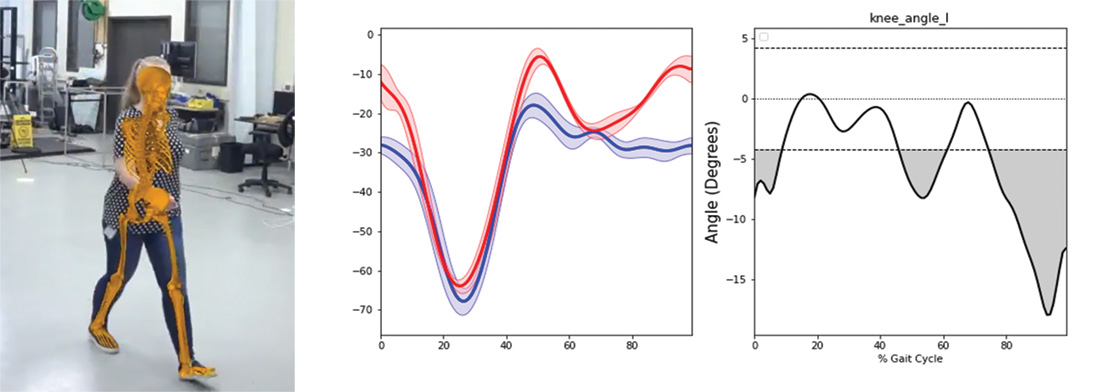
Using the ENABLE markerless biomechanics system, SwRI can accurately and automatically quantify a person’s detailed 3D movement from video. This comprehensive full-body gait analysis can capture kinematic variables in the hips, knees, ankles, shoulders, elbows, etc.